

Summer Research in the Physics Department has started. The largest group of students will begin their summers of work next week, but there are a few who started this week or earlier.
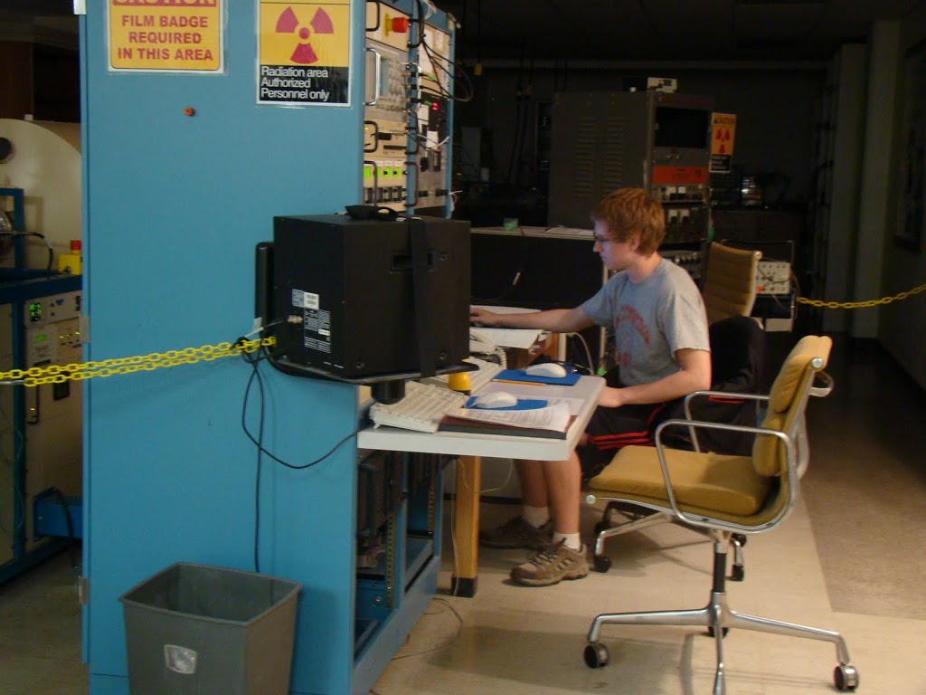 This afternoon, Kevin Krueger, who is working for Graham Peaslee (Chemistry) and Paul DeYoung, was running the accelerator at HIBAL in the basement of VanderWerf Hall.
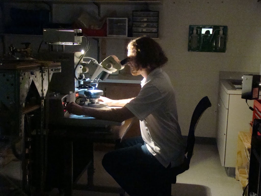
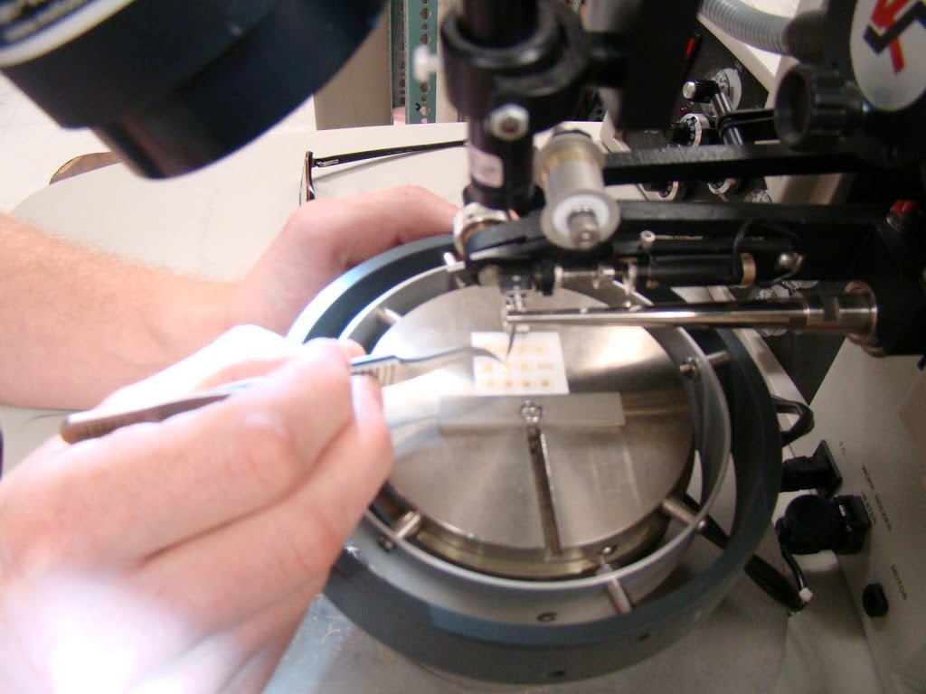
Down the hall in the Microwave Lab, Andrew Bunnell, who is working for Steve Remillard, was using the wire bonder.
We look forward to many more students joining us next week and the weeks following.


